
Thursday, 28 March 2013
Wednesday, 27 March 2013
MAJOR CAUSES OF VISUAL IMPAIRMENT IN INFANTS AND TODDLERS
|
There are many possible defects or diseases of the visual system , but,
fortunately, many of them appear after the first few years
of life. There are still many defects, diseases, infections, disorders and malformations that can affect the visual system in infants and toddlers. Only a few of the
many visual disorders found in young children is described below:
Cataracts: defined as a clouding of the lens of the eye;
can be congenital, caused by trauma, or associated with disease; when caused
by maternal rubella, cataracts are not removed early, and
acuity never develops well; if not caused by rubella,
cataracts are surgically removed soon after birth (usually within the first
two months), to allow the retina to be stimulated by light within the first
6-8 weeks of life; good acuity is possible if cataracts are removed early
enough.
Glaucoma- (infantile): (also known as
"buphthalmos") intraocular pressure build-up caused by an imbalance
between the rate of production of the aqueous fluid and the rate of normal
drainage; must be treated medically (often surgically).
Cortical Visual Impairment (CVI): apparent lack of or
reduction in vision when eyes appear to be normal; cause of the visual
reduction is in the visual cortex of the brain; there is no nystagmus;
special intervention techniques are indicated (contact VI teacher).
Infections: many types, with a variety of symptoms; most
common involve the conjunctiva (thin layer of tissue lining the eyelids and
connected to top layer of sclera); require medical treatment (usually
medication); other systemic infections (toxoplasmosis, herpes,
cytomegalovirus) can also involve the visual system.
Malformations: many types; most common are clefts in the
iris, dislocated lens, and syndrome-related abnormalities; may have prenatal
causes
Ocular-muscle problems: most common is strabismus
(one or both eyes out of alignment); can be outward, inward, upward, or
downward, depending on which muscle(s) are affected; must be evaluated
medically, for possible surgical treatment; if noticed after 6 months of age,
child should be seen by an eye specialist; treatment can be before the child
is a year old; every year of delay past age two lessens the chances for good
prognosis in acuity; can cause loss of or diminished acuity in one eye
(amblyopia) if not treated.
Nystagmus is another ocular-muscle anomaly; manifested by
involuntary eye movements, usually noted as "jerky" or
"jumpy" eye movement; occasionally occurs alone but most often
accompanies other eye conditions; there is no cure; acuity may be reduced,
but visual function may improve with age.
Ocular trauma: occurs when the eyeball is hit, lacerated,
or punctured; always requires medical evaluation and treatment.
Optic nerve defects: Optic atrophy
occurs when, for a number of possible reasons, the optic nerve does not
function properly; may result in inconsistent visual functioning; often
causes reduced acuity; there are usually no outward indicators - the eyes
appear normal ; glasses will not improve acuity; must be medically diagnosed;
the phrase pale optic disk(s) suggests the possibility of optic atrophy. Optic
nerve hypoplasia (ONH) differs from optic atrophy; in ONH, the optic
nerve has regressed in development (usually during the prenatal period, and
usually caused by a prenatal insult to the neurological system); must be
medically diagnose; may have accompanying brain malformation and/or endocrine
problems; there is no treatment, and glasses will not help. Septo-optic
dysplasia seem to be an extreme form of ONH.
Refractive errors: (nearsightedness, farsightedness,
astigmatism) These are the only defects glasses will help, but, since the
infant eye is still developing (and clear acuity is still poor), they are
usually not identified as problems in the early months. If present to a
marked degree after about 12 months, they may require a
prescription for glasses but most toddlers will not need corrective lenses.
If acuity seems to be reduced (not within normal ranges) after about age 2,
medical evaluation is recommended. In the case of premature infants, an eye
specialist should monitor vision periodically from birth.
Retinoblastoma: a tumor behind the eye which, if left
untreated, can be both blinding and life-threatening; medical treatment
(chemotherapy and/or enucleation) is essential, usually before age 2.
Retinopathy of Prematurity (ROP) : (formerly called
retrolental fibroplasia, or RLF) a condition found primarily (but not
exclusively) among premature infants; despite the suspected role of oxygen in
this disease, prematurity seems to be the major factor; identified medically;
cryotherapy appears to halt the progression of the disease; visual function
can range from near normal acuity to total blindness, depending on the stage
of the disease; about a fourth of children with ROP have severe visual
impairment; many of these children are also myopic (nearsighted).
NORMAL DEVELOPMENT
|
||||||||||||||||||||||||||||||||||||||||||||||||||||||||||||||||||||||||||||||||||||||||||
MONITORING VISUAL DEVELOPMENT "FROM BIRTH TO THREE YEARS OF AGE"
In the early months of life, the visual
system is still maturing; it is not fully developed at birth (and is even
less developed in the premature infant). From birth to maturity, the eye
increases to three times its size at birth, and most of this growth is
complete by age 3; one third of the eye's growth in diameter is in the first
year of life. Some knowledge of normal visual development is necessary if
abnormalities are to be noted. The following information gives indicators of
normal visual development in young children from birth to three years.
In a
premature infant: (depending on the extent of prematurity)
The eyelids may not have fully separated; the iris may not constrict or
dilate; the aqueous drainage system may not be fully functional; the choroid
may lack pigment; retinal blood vessel's may be immature; optic nerve fibers
may not be myelinized; there may still be a Pupillary membrane and/or a
hyaloid system. Functional implications: lack of ability to control light
entering the eye; visual system is not ready to function.
.
At birth: The irises of Caucasian infants may have a gray
or bluish appearance; natural color develops as pigment forms. The eyes'
pupils are not able to dilate fully yet. The curvature of the lens is nearly
spherical. The retina (especially the macula) is not fully developed. The
infant is moderately farsighted and has some degree of astigmatism.
Functional implications: The newborn has poor fixation ability, a very
limited ability to discriminate color, limited visual fields, and an
estimated visual acuity of somewhere between 20/200 and 20/400.
By 1 month: The infant can follow a slowly moving black and
white target intermittently to midline; he/she will blink at a light flash,
may also intermittently follow faces (usually with the eyes and head both
moving together). Acuity is still poor (in the 20/200 to 20/400 range), and
ocular movements may often be uncoordinated. There is a preference for black
and white designs, especially checkerboards and designs with angles.
By 2 months: Brief fixation occurs sporadically, although
ocular movements may still be uncoordinated; there may be attention to
objects up to 6' away. The infant may follow
vertical movements better than horizontal , and is beginning to be aware of
colors (primarily red and yellow). There is probably still a preference for
black and white designs.

By 3 months: Ocular movements are coordinated most of the
time; attraction is to both black and white and colored (yellow and red)
targets. The infant is capable of glancing at smaller targets (as small as
1"), and is interested in faces; visual attention and visual searching
begins. The infant begins to associate visual stimuli and an event (e.g., the
bottle and feeding).
By 4 months: "Hand regard" occurs at about 15 weeks;
there is marked interest in the infant's own hands. He/she is beginning to
shift gaze, and reacts (usually smiles) to familiar faces. He/she is able to
follow a visual target the size of a finger puppet past midline, and can
track horizontally, vertically, and in a circle. Visual acuity may be in the
20/200 to 20/300 range.

By 5 months: The infant is able to look at (visually examine)
an object in his/her own hands; ocular movement although still uncoordinated
at times, is smoother. The infant is visually aware of the environment
("explores" visually), and can shift gaze from near to far easily;
he/she can "study" objects visually at near point, and can converge
the eyes to do so; can fixate at 3'. Eye-hand coordination (reach) is usually
achieved by now.

By 6 months: Acuity is 20/200 or better, but eye movements are
coordinated and smooth; vision can be used efficiently at both near point and
distance. The child recognizes and differentiates faces at 6', and can reach
for and grasp a visual target. Hand movements are monitored visually; has
visually directed reach." May be interested in watching falling objects,
and usually fixates on where the object disappears.
Between 6 and 9 months: Acuity improves rapidly (to near normal);
"explores" visually (examines objects in hands visually, and
watches what is going on around him/her). Can transfer objects from hand to
hand, and may be interested in geometric patterns.

Between 9 months and a year: The child can visually spot a small (2-3mm)
object nearby; watches faces and tries to imitate expressions; searches for
hidden objects after observing the "hiding;" visually alert to new
people, objects, surroundings; can differentiate between known and unfamiliar
people; vision motivates and monitors movement towards a desired object.

By 1 year: Both near and distant acuities are good (in the
20/50 range); there may be some mild farsightedness, but there is ability to
focus, accommodate (shift between far and near vision tasks), and the child
has depth perception; he/she can discriminate between simple geometric forms
(circle, triangle, square), scribbles with a crayon, and is visually
interested in pictures. Vision lures the child into the environment. Can
track across a 180 degree arc.

By 2 years: Myelinization of the optic nerve is completed.
There is vertical (upright) orientation; all optical skills are smooth and
well coordinated. Acuity is 20/20 to 20/30 (normal). The child can imitate
movements, can match same objects by single properties (color, shape), arid
can point to specific pictures in a book.
By 3 years: Retinal tissue is mature. The child can complete a
simple formboard correctly (based on visual memory), can do simple puzzles,
can draw a crude circle, and can put 1" pegs into holes.
 |
Tuesday, 26 March 2013
Farsighted engineer invents bionic eye to help the blind
For
UCLA bioengineering professor Wentai Liu, more than two decades of visionary
research burst into the headlines last month when the FDA approved what it
called “the first bionic eye for the blind.”
The Argus II Retinal Prosthesis System — developed by a team of physicians and engineers from around the country — aids adults who have lost their eyesight due to retinitis pigmentosa (RP), age-related macular degeneration or other eye diseases that destroy the retina’s light-sensitive photoreceptors.
At the heart of the device is a tiny yet powerful computer chip developed by Liu that, when implanted in the retina, effectively sidesteps the damaged photoreceptors to “trick” the eye into seeing. The Argus II operates with a miniature video camera mounted on a pair of eyeglasses that sends information about images it detects to a microprocessor worn on the user’s waistband. The microprocessor wirelessly transmits electronic signals to the computer chip, a fingernail-size grid made up of 60 circuits. These chips stimulate the retina’s nerve cells with electronic impulses which head up the optic nerve to the brain’s visual cortex. There, the brain assembles them into a composite image.
Recipients of the retinal implant can read oversized letters of the alphabet, discern objects and movement, and even see the outlines and some details of faces. And while the picture is far from perfect — the healthy human eye sees at a much higher resolution — it’s a breakthrough for people like the first patient, a man in his 70s who was blinded at age 20 by RP, to receive the implant in clinical trials. “It was the first time he’d seen light in a half-century,” said Liu, adding that “it feels good as the engineer” to have helped make this possible.

Liu joined the Artificial Retina Project in 1988 as a professor of computer and electrical engineering at North Carolina State University. The multidisciplinary research project was funded by the U.S. Department of Energy’s Office of Science because it envisioned a potential pandemic of eyesight loss in America’s aging population. Leading the project was Duke University ophthalmologist and neurosurgeon Dr. Mark Humayun, now on faculty at USC. He tapped Liu to engineer the artificial retina.
“I thought it was a great idea,” Liu said. “But I asked, ‘What can I do?’ because I didn’t know much about biology.” Humayun handed him a six-inch-thick medical manual on the retina. “The learning curve was very steep,” Liu recalled with a laugh.
However, Liu’s fellow engineers questioned his sanity. “I was working on integrated chip design and had just gotten tenure when I signed on to this project. They said, ‘You’re crazy!’ But I’m glad I made that choice, getting into this new field.”
Wednesday, 13 March 2013
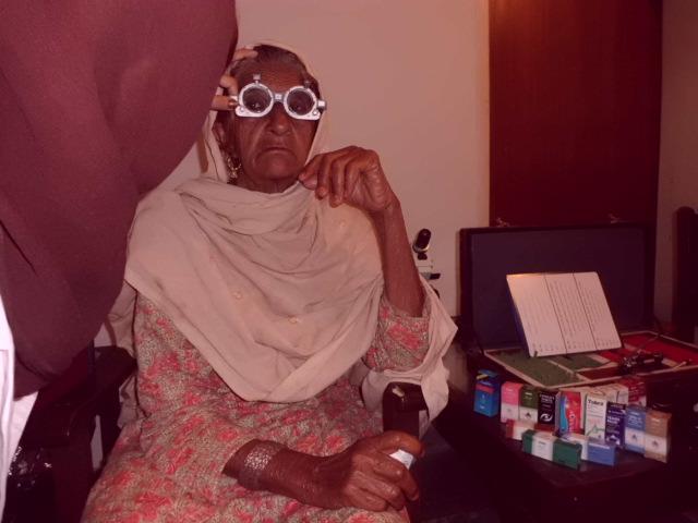
Free Eye Camp "10 March 2013" - POS PUNJAB
Highlights of the free eye camp held on 10 march 2013 by Pakistan Optometric Society. A Total number of 273 patients screened and dispensed medicines. 127 refractions performed, Free of cost spectacles were provided. A number of 39 cataracts booked and refereed for IOL Implantations.
Optometrists: Aamer Niazi, Lubna Iram, Miss Sadia.
Camp Review in pictures :-


Subscribe to:
Posts (Atom)